Figure 1.

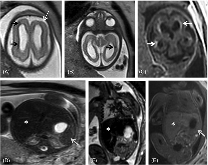
Fetal magnetic resonance imaging (MRI) at 29 gestational weeks referred for microcephaly and splenomegaly. There is parenchymal loss, with global thinning of the cerebral mantle and consequent bilateral ventriculomegaly. Small areas of focal signal anomaly can be detected on T2WI (A, B, black arrows), as well as frontal polymicrogyria (A, white dashed arrow). Calcifications can be identified on T1WI (c, white arrows). Enlarged spleen (gray arrow) and liver (asterisk) can be identified on T2W steady state free precession (D), echo planar imaging (E) and T1W images (F), with slight signal intensity anomaly on the latter
