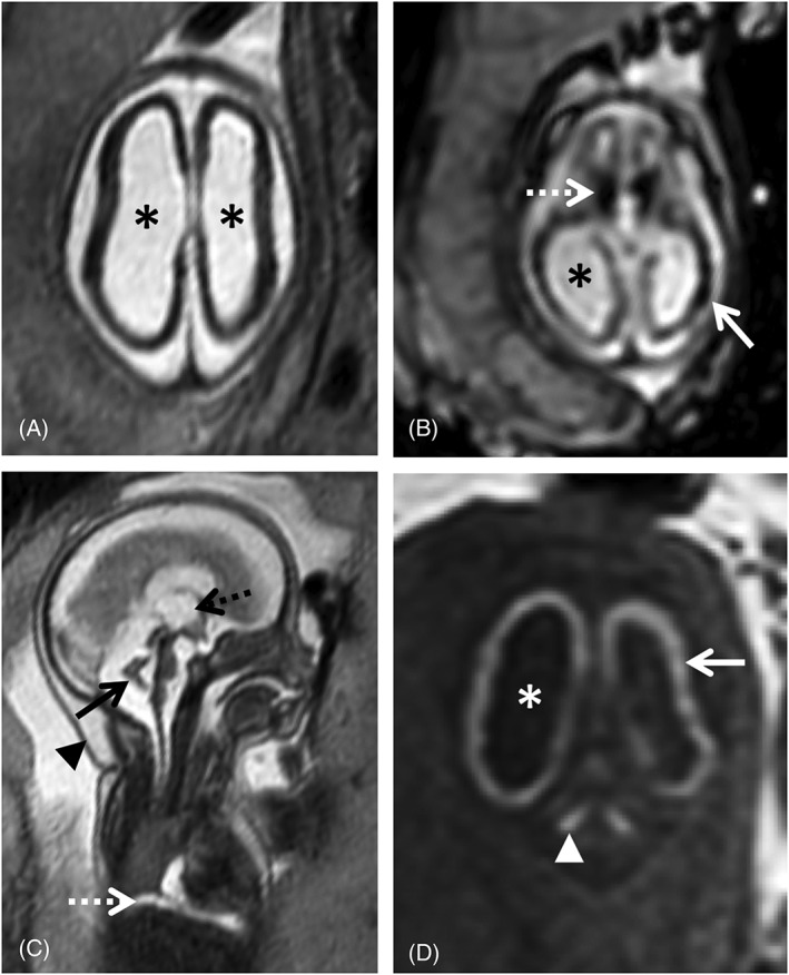
Figure 2.

24 gestational weeks fetus referred for fetal magnetic resonance imaging (MRI) for suspected lissencephaly and cerebellar hypoplasia. T2w single shot fast spin echo axial (A) and sagittal (C), axial echo planar imaging (EPI) (B) and coronal T1WI (D) show a marked reduction of the cerebral parenchyma thickness, with diffuse low SI on T2WI (A) and severe ventriculomegaly (asterisk), including dilatation of the third ventricle (C, black dashed arrow). There is diffuse high SI on T1WI of the supratentorial parenchyma (D, white arrow) compatible with presence of calcifications, confirmed on EP images (B) and particularly evident in the basal ganglia (B, white dashed arrow). There is cerebellar hypoplasia (C, black arrow) associated with deep gray nuclei calcifications (D, white arrowhead). Furthermore, small pleural effusion can be seen on sagittal T2WI (C, white dashed arrow). Findings were confirmed on postmortem MRI (not shown). Additionally, there is skin edema/thickening of the skin (C, black arrow head)
