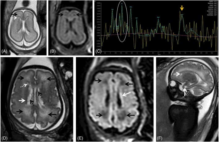
Figure 3.

Fetal magnetic resonance imaging at 24 gestational weeks (GW) referred for maternal cytomegalovirus seroconversion and normal brain ultrasound. On T2 weighted images (WI) slight periventricular caps can be identified frontally (A, black arrow) as well as a small periventricular cyst. PRESS spectroscopy performed with a short TE (35 ms) depicts a myoinositol peak (myo‐inositol, white circle, C) and a lactate peak (Lac, yellow arrow, C), raising the suspicion of more extensive brain involvement. Follow up at 34 gestational weeks shows progression of white matter signal changes in the frontal and parieto‐occipital regions bilaterally (black arrows, D) that also have translation on the FLAIR image (black arrows, E). The presence of the small periventricular cyst can be confirmed (D, F, white dashed arrow) and there is the additional finding of a temporal pole cyst (F, white arrow) and irregularity and signal alteration of the ventricular lining (hypointense on T2WI, D, white arrow; and hyperintense on t2W FLAIR images, E, white arrow) with intraventricular septations/pseudocysts (D, black dashed arrow)
