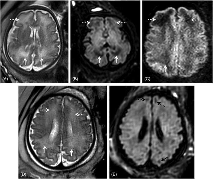
Figure 4.

Two fetuses referred for fetal magnetic resonance imaging at 34 gestational weeks for congenital cytomegalovirus (cCMV) infection (A‐C) and abdominal cyst (D‐E). White matter hyperintensities can be identified on T2WI in the frontal (A, D, white dashed arrows) and parietal‐occipital parieto‐occipital (A, D, white arrows) regions. On T2w echo planar imaging‐FLAIR images (B, E) there is corresponding hypointensity in the cCMV patient (B, white/dashed arrows), as well as low SI on the zoom diffusion weighted image (C, white/dashed arrows), but not the control (E) except in the expected gyral crests corresponding to remnants of the subplate (E, black dashed and full arrows)
