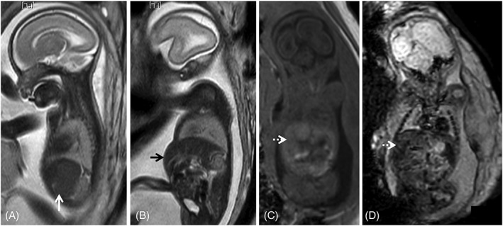
Figure 7.

Fetal magnetic resonance imaging of a cytomegalovirus (CMV) positive fetus at 24 gestational weeks. T2WI depict splenomegaly (A, white arrow) and hepatomegaly (B, black arrow), protruding in the anterior abdominal wall. There is associated abnormal liver signal: isointense on T1WI (C, dashed white arrow) and on echo planar images (D, dashed white arrow), which cannot be identified on T2WI. These findings are often found in fetal infections and may help guide the diagnosis when brain anomalies are found, in the absence of a definite diagnosis of CMV infection
