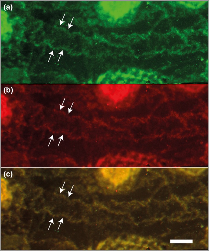
Figure 2.

Patient IgG binds to deposited laminin‐332. Double staining of living cells incubated with antilaminin‐332 patient IgG for (a) human IgG and (b) the laminin‐332 γ2 subunit shows complete colocalization in the merged panel (c). The white bar represents 50 μm.
