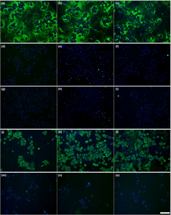
Figure 3.

IgG binding patterns after incubation of different types of pemphigoid sera with living keratinocytes. Binding patterns after 1‐h incubation with sera of patients with (a–c) antilaminin‐332 mucous membrane pemphigoid, (d–f) epidermolysis bullosa acquisita, (g–i) anti‐p200 pemphigoid and (j–l) bullous pemphigoid and (m–o) normal human control sera. The white bar represents 150 μm.
