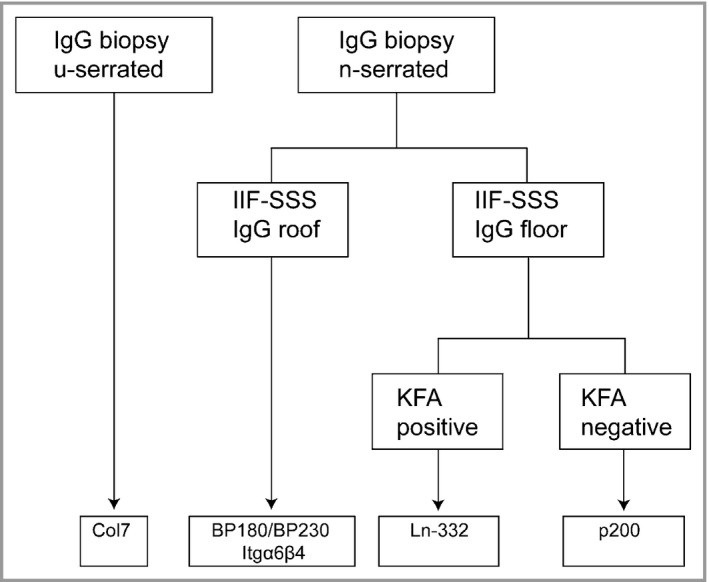
Figure 5.

Flowchart for the diagnosis of different subtypes of pemphigoid diseases. By combining (i) the serration pattern of basement membrane zone deposition in the skin, (ii) indirect immunofluorescence (IIF) of serum on salt‐split skin (SSS) and (iii) the keratinocyte footprint assay (KFA), rapid identification of the antigens recognized can be achieved. Col7, type VII collagen; Itgα6β4, integrin α6β4; Ln‐332, laminin‐332.
