Figure 3.

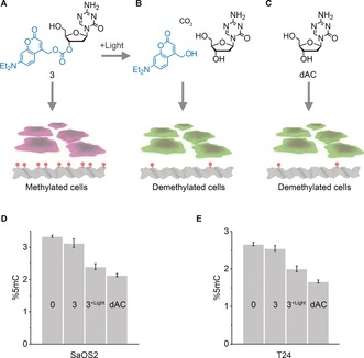
DEACMOC‐dAC 3 can be photo‐deprotected to re‐activate its inhibitory effect on DNMT and lower DNA methylation levels in cells. a–c) Schematic of cell treatment conditions and expected qualitative changes in DNA methylation levels. Treatment with 3 in the absence of light maintains high methylation levels (a), while illumination restores dAC activity to lower DNA methylation (b) to levels close to unmodified dAC (c). The concentration of 3 and dAC was 0.1 μm. Cells take up photocaged dAC at up to 4.5 μm within 1 h as shown using cell viability read‐out. d,e) Treatment‐dependent changes in methylation levels in SaOS2 (d) and T24 cell lines (e) for condition in (a–c) and 0 μm dAC under light exposure, as quantified by LC‐MS. DNA methylation levels (%5mC) are expressed as a percentage of total cytosines and analysed in biological triplicates.
