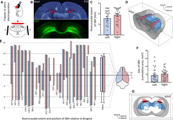
Figure 1.

Anatomical features of bilateral Dcx‐knockdown (KD) rats with bilateral subcortical band heterotopia. A, A schematic view of the tripolar in utero electroporation procedure, showing the placement of tweezer‐type electrodes (black, negative pole) laterally pinching the embryo’s head, and that of the third electrode (red, positive pole), placed rostrally and medially. The electric field induced by the potential applied to the electrodes enables a simultaneous and bilateral electroporation of cortical progenitors lining the lateral ventricles (gray area and arrows). B, Bright‐field and fluorescent microphotographs of a postnatal day 15 (P15) neocortical section from a bilateral Dcx‐KD rat showing bilateral subcortical band heterotopia (SBH; red asterisks), mostly composed of green fluorescent protein (GFP)‐expressing neurons. Bundles of GFP‐expressing callosal axons are also visible. C, Bar graphs showing the rostrocaudal extent of SBH in the left (blue) and right (pink) hemispheres of 25 adult bilateral Dcx‐KD rats. D, Dorsolateral view of the rostral part of a three‐dimensionally reconstructed adult brain with bilateral SBH (in red). The white matter (wm) is delineated in blue. E, Clustered bar graphs showing the rostrocaudal extent and position of SBH relative to bregma in the 25 Dcx‐KD rats analyzed, in the left (blue) and right (pink) hemispheres. Animal identifier codes are given below each pairs of bars. F, Bar graphs showing the size of SBH (depicted as cumulative area) in the left (blue) and right (pink) hemispheres of 25 adult bilateral Dcx‐KD rats. Open circles in C and F correspond to values for individual rats. G, Rear view of the same brain as in D
