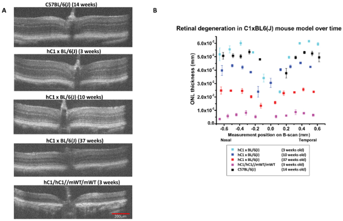
Figure 2: Progressive Outer Retinal Degeneration in partially humanized adRP model as measured by HR-SD-OCT.
(A) HR-SD-OCT images of the retina were obtained for the adRP model (hC1 x BL/6 (J)) at different ages, the C57BL/6(J) control at 14 weeks, and the homozygous hC1 mutant line at 3 weeks. The outer retina had a normal appearance at 3 weeks in the adRP model, but there was evidence of progressive ONL thinning and disorganization at and beyond 10 weeks of age. By 37 weeks, the (hC1 x BL/6(J)) demonstrated extensive outer retinal degeneration. All OCT scans were in vicinity of optic nerve. (B) The ONL thickness (in mm) along the horizontal axis through the optic nerve was plotted for control (C57BL/6(J)), hC1, and adRP animals of differing ages. There was progressive loss of ONL thickness in the partially humanized adRP model. ONL loss was greater than 60% by 37 weeks of age. Error bars = standard error of the mean. Red scale bar = 200 μm and all the images are the same scale.

