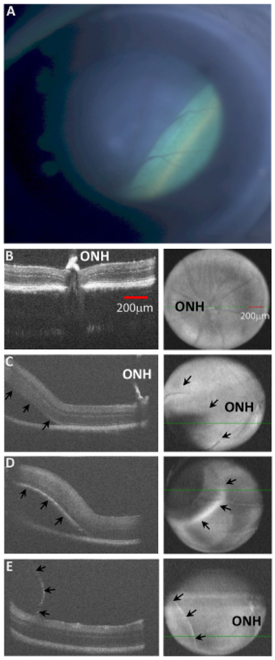
Figure 4: Types of intraocular Injections in mice identified by HR-SD-OCT.

(A). A (hC1xBL/6(J)) mouse was injected with ~1 μL of fluid via Inferonasal transcleral transchoroidal injection. The resulting retinal detachment is seen as the green lower right region of the en face image, creating a sharp border at the leading edge of the bleb on the right side of the image. The OCT fundus image of an injection site only exhibits subtle differences depending upon the position of the fluid filled cavity, because the image is a compilation of all the b-scans from the entire retinal thickness. In addition, the injection bleb changes the distance of the retinal surface from the OCT headpiece, producing an unfocused region at the injection site. (B) The OCT b-scan of a non-injected retina is demonstrated. The ONH is labeled. (C) A subretinal injection is demonstrated. The ONH is labeled, and arrows in the en face image (right panels) show the posterior border of the detachment. (D) A choroidal injection is demonstrated with a clear elevation (upward displacement) of the RPE layer (hyper-reflective curve) of the lower border of the retina (arrows) and significant loss of hyper-reflectivity of the RPE and choroid layers below the injected fluid. Compare arrows in the images (C vs. D). (E) A retinal schisis is demonstrated near the nerve fiber layer. Observe the very thin hyper-reflective membrane encapsulating the injected fluid, while the retina remains attached to the RPE. Subtle differences between the three different detachments can also be visualized in the en face images (C, D, and E). The subretinal detachment has a border which is difficult to visualize (arrows in C), while the choroidal injection creates a blurred hyper reflective rim at the leading edge of the bleb, and the retinal schisis is evident by the sharp demarcation of its leading edge (E). Both red scale bars = 200 μm in 4B. All images 4B through 4E are scaled equally.
