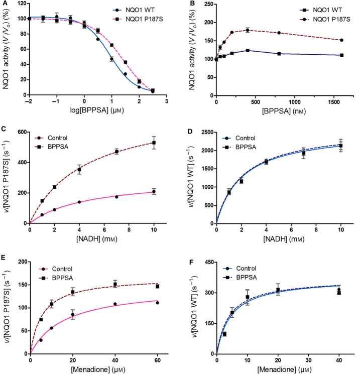
Figure 7.

Inhibition, activation, and steady‐state kinetic assays for NQO1 WT and the P187S variant in the presence and absence of BPPSA. Inhibition by BPPSA is (A) shown for NQO1 WT (in blue) and the NQO1 P187S variant (in pink) and the corresponding IC50 values were calculated to be 8.6 ± 1.1 μm for NQO1 WT and 26.2 ± 1.2 μm for the NQO1 P187S variant. (B) Activation of NQO1 WT (dark blue) and the NQO1 P187S variant (maroon) in the presence of BPPSA. The velocity v over the enzyme concentration is plotted against the concentration of NADH for (C) the NQO1 P187S variant and (D) NQO1 WT and against the concentration of menadione for (E) the NQO1 P187S variant and (F) NQO1 WT.
