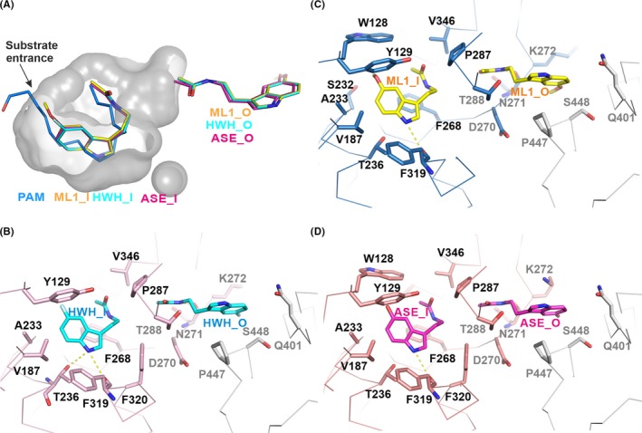
Figure 3.

Notum enzymatic pocket and inhibitor binding details. A, The enzyme pocket is shown as a grey surface with 50% transparency. The bound ML1 (yellow; PDB code: 6TR5), HWH (cyan; PDB code: 6TR7) and ASE (magenta; PDB code: 6TR6) and palmitoleate (blue; PDB code 4UZQ) are shown as sticks. The outside pocket inhibitors are also shown. B‐D, The binding details of the Notum inhibitors. The Notum structure is shown as a ribbon with a symmetry‐related molecule in grey. The Notum residues interacting with enzymatic pocket inhibitors are labelled in black while the outside ones are in grey. Hydrogen bonds are shown as dashed yellow lines
