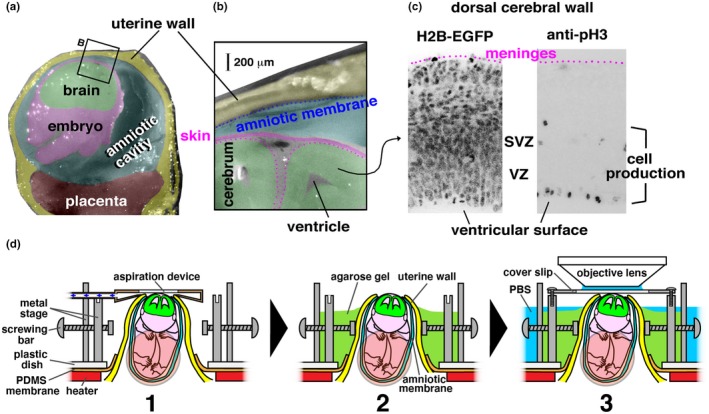
Figure 1.

Objective and methods of in utero two‐photon microscopic live observation. (a) Cross‐section photomicrograph of the uterus and the amniotic cavity containing an E14 mouse embryo (cut was coronal at the head of the embryo and through the placenta). (b) Magnified view of the uterine wall, the amniotic membrane, the amniotic cavity, and the dorsal portion of the embryo's head. (c) Photomicrographs of the dorsal cerebral wall where either the H2B‐EGFP signal (all cell nuclei and chromosomal condensation during M phase) was observed live or M‐phase cells were visualized immunohistochemically with anti‐phosphohistone H3 (pH3). VZ, ventricular zone. SVZ, subventricular zone. (d) Steps (1–3) and devices for posturing and holding in utero growing embryos are shown
