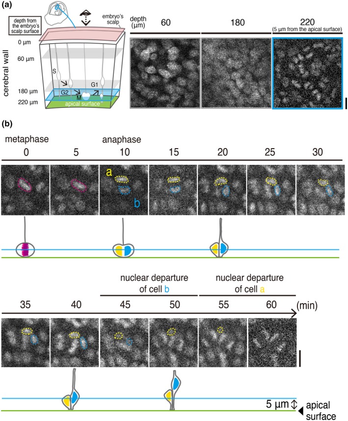
Figure 2.

In utero 2PM view of an E13 H2B‐EGFP transgenic mouse embryo enables observation of cell‐production behaviors by NPCs near the apical surface of the cerebral wall. (a) Schematic display of our 2PM scanning procedures until reaching 5 μm from the apical surface; the accompanying obtained H2B‐EGFP images are shown (see also Movie S1). (b) An example of time‐lapse monitoring of the division of an NPC and the IKNM exhibited by its daughter cells. Magenta circle, metaphase plate (chromosomal condensation) of a mitotic mother cell. Yellow and cyan circles, daughter cells' condensed chromosomes and nuclei. See also Movie S2. Scale, 10 μm
