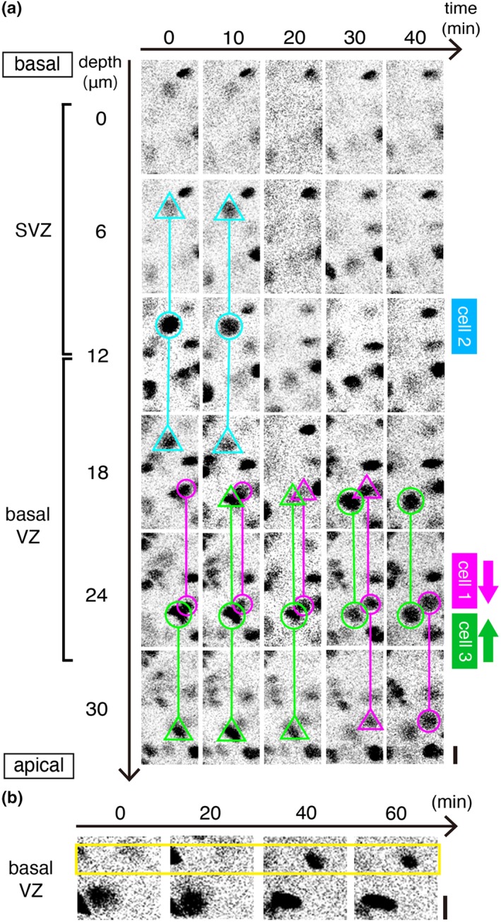
Figure 5.

Intravital 2PM that captured cell‐cycle‐associated events including the extinction and the emergence of mAG signal in Fucci embryos in utero. (a) A time series showing cell‐cycle‐associated NPC dynamics in the SVZ and the basal VZ of an E14 Fucci (S/G2/M‐Green) embryo. Scanning was performed from the SVZ (0 μm) to the middle VZ (30 μm). Cell 1 (magenta) showed apical‐ward nuclear migration (corresponding to “event 1” of Figure 4a), as also shown in Figure 4b, typical to G2‐phase NPCs. Cell 2 (light blue) was initially brightly mAG+, large in size and round in shape, and its mAG then became undetectable, typical phenomenon in M‐phase‐progressing (dividing) cells (corresponding to “event 2” of Figure 4a). Cell 3 (green) exhibited basal‐ward movement (corresponding to “event 3” of Figure 4a), a phenomenon preparative for non‐surface mitosis. Scale, 10 μm. (b) A time series showing emergence of a new mAG+ nucleus in the basal VZ, indicative of either chronological entrance of a G1‐finishing NPC into S phase (corresponding to “event 4” of Figure 4a) or spatial entrance of an S‐phase NPC nucleus into the basal VZ. Scale, 10 μm
