Figure 1.

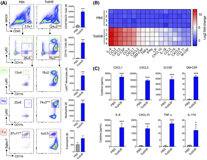
Intrarectal instillation of C difficile toxins TcdA and TcdB (TcdA/B) leads to the robust influx of innate immune cells into the colonic lamina propria and increased colonic production of inflammatory mediators. TcdA/B were intrarectally administered for 4 h (25 μg in PBS; PBS alone in control group), after which cells from the colonic lamina propria were isolated and stained for cell surface markers. A, Stained cells were fractioned based on the expression of CD45 and MHCII. CD45+MHCII− cells were further fractioned into CD11b+ cells with varying levels of Ly6C expression. These CD11b+ cells were composed of Ly6Chi inflammatory monocytes (green box, MΦ), Ly6CintLy6G+ neutrophils (blue box, NΦ), and Ly6CloSiglec‐F+ eosinophils (red box, MΦ). Cell percentage of each population is shown in the corresponding representative flow cytometry plot, and total cell count is depicted in the accompanying bar plot. B, Colonic tissue homogenates from control (PBS‐treated)‐ and TcdA/B‐treated mice were assessed for cytokine production using the Luminex discovery assay. The heatmap in panel B shows the top colonic cytokines that are differentially produced between PBS‐ and TcdA/B‐treated mice (Log2 fold change in TcdA/B vs PBS). C, Protein levels of the top cytokines upregulated in the colon of TcdA/B‐treated mice and play a role in the pathogenesis of C difficile infections (n = 4 per group). All data are expressed as SEM. Student's t test, *P < .05, ***P < .005
