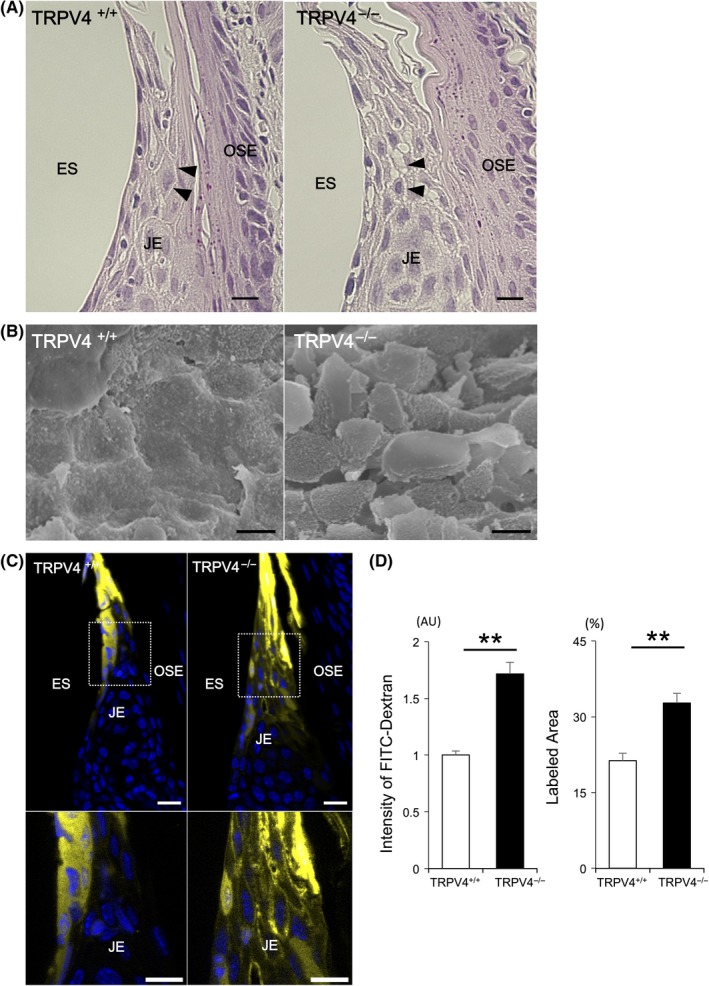
Figure 2.

Morphological characteristics and permeability of the JE. A, Light micrographs of hematoxylin and eosin staining of the JE. No apparent structural differences in the JE are observed between TRPV4+/+ and TRPV4−/− mice. Note the wider intercellular spaces (arrowheads) in TRPV4−/− mice compared with TRPV4+/+ mice. B, Scanning electron micrographs of the junctional epithelial surface in TRPV4+/+ and TRPV4−/− mice. Rough epithelial arrangement of the junctional epithelial surface is observed in TRPV4−/− mice compared with TRPV4+/+ mice. An apparent polygonal cell shape is seen in TRPV4−/− mice. C, Permeability assay of the JE. The coronal JE was labeled with fluorescent dextran. Dextran is observed at higher levels in the intercellular spaces of the coronal JE in TRPV4−/− mice compared with TRPV4+/+ mice. D, The intensity in the JE and ratio of labeled area are significantly higher in TRPV4−/− mice than in TRPV4+/+ mice. All data are expressed as mean ± SE (n = 4). **P < .01. ES, enamel space; JE, junctional epithelium; OE, oral epithelium; OSE, oral sulcular epithelium. Scale bars: 10 µm
