Figure 3.

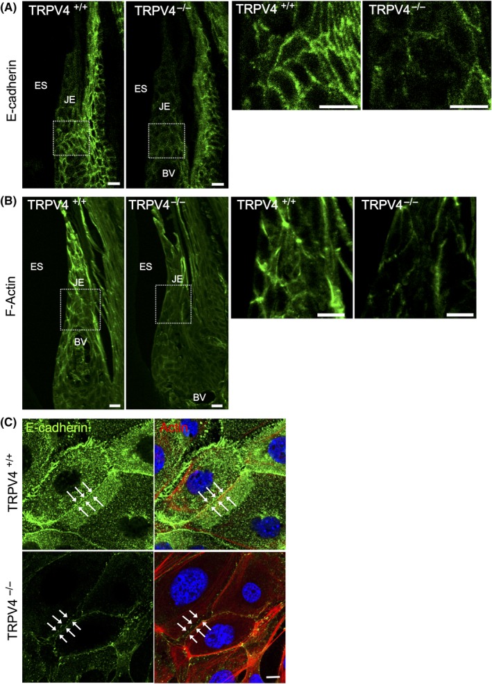
Effects of TRPV4 on the distribution of adherens junction proteins. A, E‐cadherin immunostaining in the JE has a mesh‐like pattern depicting an intercellular arrangement. TRPV4−/− mice show weaker E‐cadherin immunoreactivity in the JE compared with TRPV4+/+ mice. B, Rhodamine‐phalloidin staining of the JE in TRPV4+/+ and TRPV4−/− mice. Peripheral actin delineates the cell surface in TRPV4+/+ mice, but is rough and weak in TRPV4−/− mice. C, Immunofluorescence photomicrographs of primary cultured oral epithelial cells from TRPV4+/+ and TRPV4−/− mice after high‐calcium medium treatment for 24 h. Representative data from one of five experiments are shown. Cells from TRPV4+/+ mice form cell‐cell contacts showing E‐cadherin immunoreactivity (green) with a linear appearance (arrows). Cells from TRPV4−/− mice have intercellular gaps with weaker wavy lines (arrows) of E‐cadherin immunoreactivity compared with cells from TRPV4+/+ mice. Punctate E‐cadherin and F‐actin staining is present in the cells from TRPV4−/− mice. Rhodamine‐phalloidin: red; DAPI: blue. BV, blood vessels; ES, enamel space; JE, junctional epithelium. Scale bars: 10 µm
