
Figure 5.
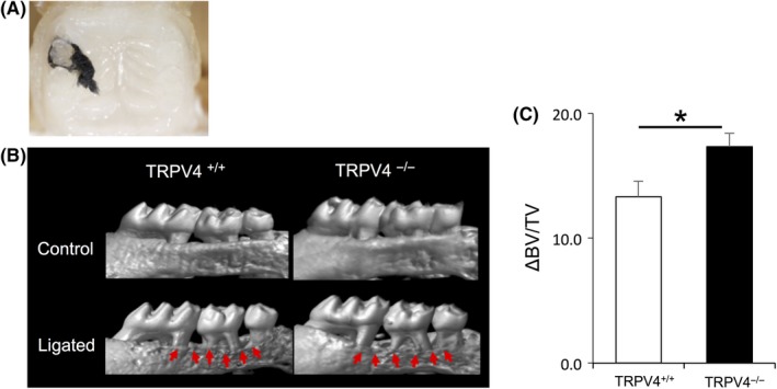
TRPV4 effect on a mouse periodontitis model. A, Representative intraoral image demonstrating a ligature placement around the second maxillary molar. B, Reconstructed micro‐CT images of the maxilla. The upper panels show no suture placement and the lower panels show suture placement. Arrows indicate areas of bone loss with increased root structure visible above the alveolar bone crest. C, Summary of the changes in bone volume per tissue volume (ΔBV/TV) at 10 d after ligature placement. Significantly larger changes are observed in TRPV4−/− mice compared with TRPV4+/+ mice. All data are expressed as mean ± SE (n = 10). *P < .05