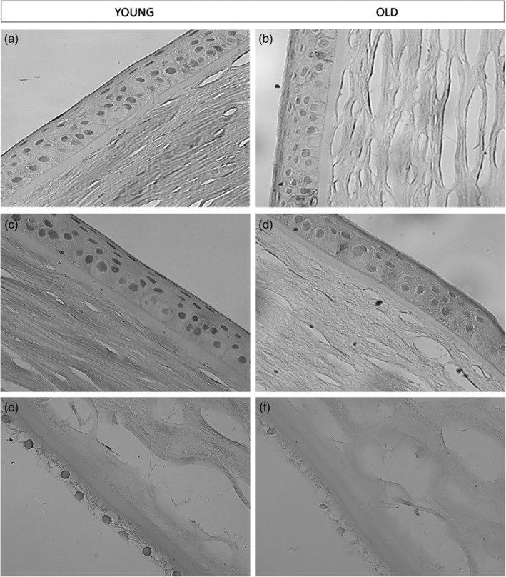
Figure 1.

Age‐related changes in the corneal layers: Light microscopy. (a) and (c) Figures show the epithelial layer of the human cornea in a young subject. We can observe a contiguous and compact paving of normal epithelial cells (×40). (b) and (d) Figures show the epithelial layer of the human cornea in an elderly subject (×40). (e) Figure shows the endothelial layer of the human cornea in a young subject (×40). (f) Figure shows the endothelial layer of the human cornea in an elderly subject. We can observe that the endothelial cells are discontinuous and partially swollen (×40).
