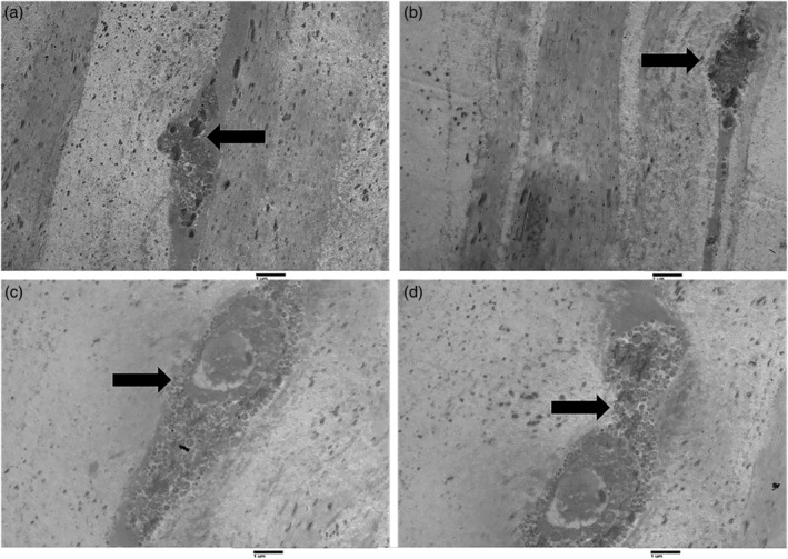
Figure 2.

Transmission electron microscopy of longitudinal sections of the human cornea in young subjects. Figures (a) and (b) show two dendrites present in the corneal stroma of young subjects (arrows). Figures (c) and (d) show a different magnification of the same structure: dendrite with a big vesicle within the corneal stroma in a young subject (arrows; magnification ×2000).
