
Figure 3.
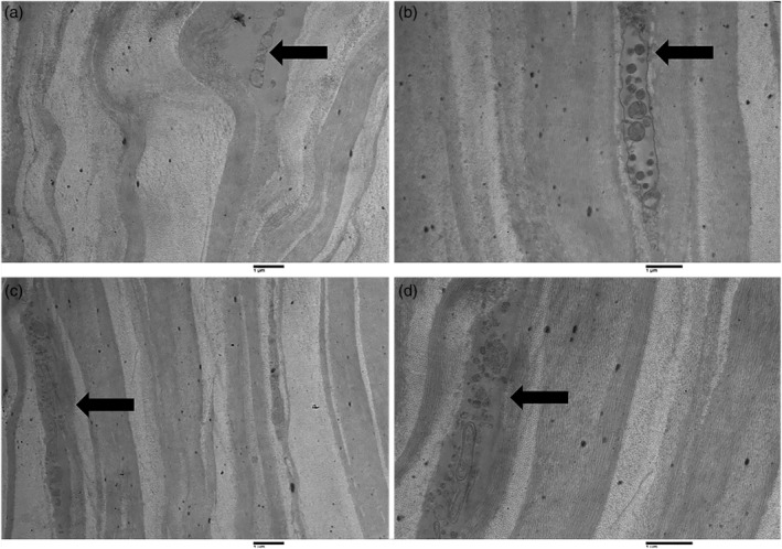
Transmission electron microscopy of longitudinal sections of the human cornea in elderly subjects. Figures (a) (magnification ×1,600) and (b) (magnification ×2,500) show two distal dendrites found in the corneal stroma of elderly subjects (arrows). Figures (c) (magnification ×1,600) and (d) (magnification ×3,150) show different magnifications of the same structure: nerve fiber, in particular, a non‐myelinated axon within the corneal stroma in an elderly subject (arrows).