Figure 1.

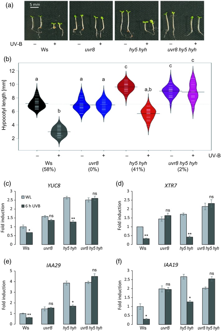
UVR8‐mediated inhibition of hypocotyl growth is partially independent on HY5 and HYH.
(a) Representative image showing the hypocotyl growth phenotype of 4‐day‐old wild‐type (Ws), uvr8‐7, hy5‐ks50 hyh‐1 and uvr8‐7 hy5‐ks50 hyh‐1 seedlings grown under white light (−UV‐B) or white light supplemented with UV‐B (+UV‐B). (b) Quantification of hypocotyl length. Beanplots represent data for n > 40 seedlings. Shared letters indicate no statistically significant difference in the means (P > 0.05). Percentages on the x‐axis indicate the relative hypocotyl growth inhibition induced by UV‐B. (c)–(f) Quantitative real‐time PCR analysis of (c) YUC8, (d) XTR7, (e) IAA29 and (f) IAA19 expression in 4‐day‐old wild‐type (Ws), uvr8‐7, hy5‐ks50 hyh‐1 and uvr8‐7 hy5‐ks50 hyh‐1 seedlings grown under white light and either exposed to narrowband UV‐B for 6 h (6 h UVB) or not (WL). Error bars represent the SE of three biological replicates. Asterisks indicate a significant decrease in transcript abundance when compared with that under white light (*P < 0.05; **P < 0.01; ns, no significant difference).
