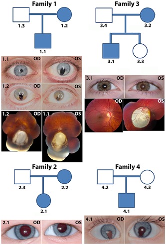
Figure 1.

Clinical findings in four coloboma families. Family 1: The proband (1.1) had bilateral coloboma in the retina and iris coloboma in the left eye; the mother (1.2) presented with left eye microcornea and posterior coloboma and right eye with chorioretinal coloboma inferior to the optic disc. Family 2: The proband (2.1) had left eye microcornea and coloboma in the optic nerve and inferior retina. Family 3: The proband (3.1) had bilateral coloboma. Family 4: The proband (4.1) had bilateral chorioretinal coloboma; the right eye was microphthalmic. OD, right eye; OS, left eye
