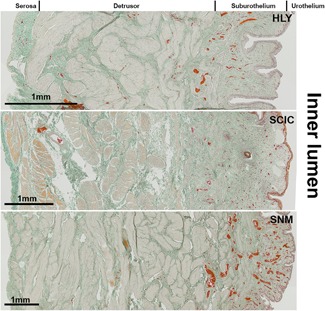
Figure 2.

Masson‐Goldner staining of the mid‐region of the urinary bladder wall of healthy reference (HLY) and SCI minipigs. The smooth muscle tissue is shown in rose and the connective tissue in green. Erythrocytes in the lumen of blood vessels are stained in red. Scale bar = 1 mm. SCI, spinal cord injury; SNM, sacral neuromodulation
