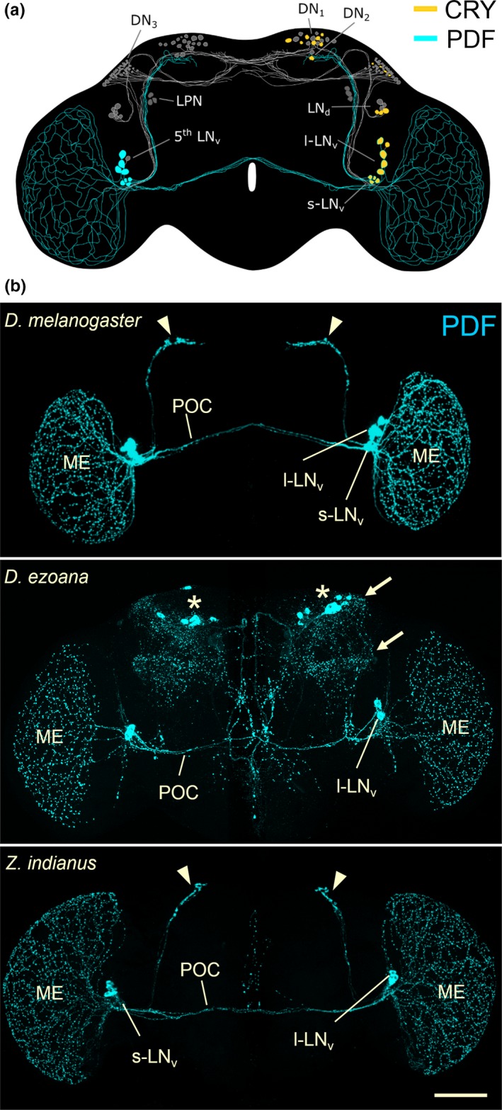
Figure 3.

Clock network in the fruit fly brain. (a) Clock neurons and their neurites in Drosophila melanogaster. The neurons consist of lateral neurons (s‐LN v, l‐LN v, LN d, 5th LN v, LPN) and dorsal neurons (DN 1, DN 2, DN 3) that are heterogeneous in respect of neuropeptide and Cryptochrome (CRY) expression. Here, we focus on the s‐LN v and l‐LN v that express the Pigment‐Dispersing Factor (PDF; cyan, left hemisphere) and CRY (yellow, right hemisphere). (b) Confocal pictures showing the PDF neurons and their arborizations in the brain and the medulla of the optic lobe for D. melanogaster, the high‐latitude species D. ezoana and the tropic species Zaprionus indianus. D. melanogaster expresses PDF in the s‐LN v and in the l‐LN v. The l‐LN v send arborizations to the medulla (ME) and via the posterior optic commissure (POC) to the other brain hemisphere. The s‐LN v project into the central brain and terminate close to the dorsal neurons (arrowheads in b, compare with a). D. ezoana expresses PDF only in the l‐LN v. These also project via the POC to the other brain hemisphere, but many fibres leave the POC and invade the central brain. Here, the latter come close to the LN d and dorsal neurons (arrows in b, compare with a). In the dorsal central brain PDF is additionally expressed in non‐clock neurons (asterisks). Z. indianus expresses PDF in the s‐LN v and the l‐LN v as does D. melanogaster and the overall arborization pattern of these two groups of neurons also largely resembles D. melanogaster. The cell bodies of the s‐LN v are only weakly stained in this individual, but the terminals of the s‐LN v in the dorsal brain are nicely visible (arrow heads). Scale bar: 100 μm
