
Figure 2. FXTAS Neuropathology.
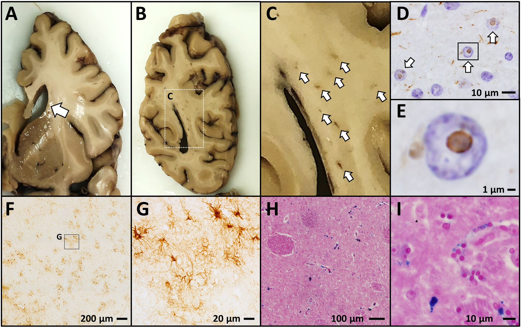
Postmortem neuropathological analysis from human FXTAS cases show a characteristic neurodegenerative phenotype which includes ventricular enlargement (A); focal white matter lesions (B,C); ubiquitinated intranuclear inclusion bodies (D,E – ubiquitin IHC with hematoxylin nuclear counterstain); patches of astrogliosis (F,G - GFAP IHC); and excessive iron accumulation (H,I – ferric iron stained using Perl’s method, Eosin counterstain).