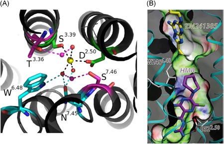
Figure 4.

A, The Na+‐distorted octahedral coordination in the A2AAR crystal structure (PDB: 4EIY): the first shell is occupied by two conserved polar residues (green) and three water molecules (small spheres), which contact with the second shell of residues (cyan), or with a second layer of water molecules connecting with a third shell of residues (magenta). B, Docking of HMA in the sodium ion binding site. The guanidinium group of HMA has a salt bridge interaction with Asp522.50 whereas the 5′‐azepane moiety of HMA clashes with Trp2466.48. ZM‐241,385 is the orthosteric antagonist. Reproduced with permission from Gutiérrez‐de Terán et al27 [Color figure can beviewed at wileyonlinelibrary.com]
