
Figure 6.
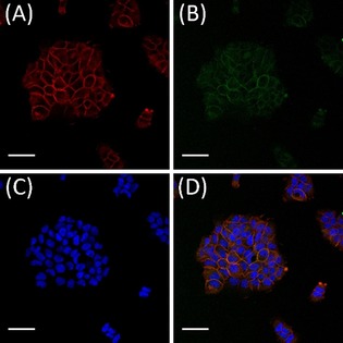
Co‐localisation of 6′ and EGFR on human epidermoid carcinoma cells. A431 cells were incubated with 100 nm 6′ for 4 h at 37 °C. Binding of the immunoconjugates to plasma membrane localised EGFR was analysed by indirect immunofluorescence. To confirm the expression of EGFR, an anti‐EGFR Alexa Fluor 647 antibody conjugate was used (A; red fluorescence). An anti‐Strep‐tag Chromeo 488 conjugate was used to detect the Strep‐tagged 6′ (B; green fluorescence). The nuclei were visualised by the DNA binding stain Hoechst 33258 (C; blue fluorescence). The overlay of (A), (B) and (C) is shown in (D). Scale bars=20 μm.