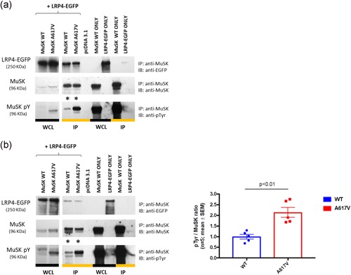
Figure 6.

Effect of p.A617V on MuSK tyrosine phosphorylation in HEK‐293T cells. (a) MuSK tyrosine phosphorylation was increased in the p.A617V variant (*) compared with WT following incubation with full‐length agrin for one hour. (b) MuSK tyrosine phosphorylation was also significantly increased in the p.A617V variant (*) in the absence of agrin incubation as shown by the densitometry analysis. Results are shown as mean ± SEM relative to WT from n = 5 experiments. Two‐tailed unpaired t test (p < .01). Lanes 1 and 2 are loading controls while lanes 5–9 are controls to ensure the specificity of the pull‐down assay. EGFP, enhanced green fluorescent protein; IP, immunoprecipitation; MuSK, muscle‐specific receptor tyrosine kinase; SEM, standard error of mean; WCL, whole cell lysates; WT, wild‐type
