
Figure 5.
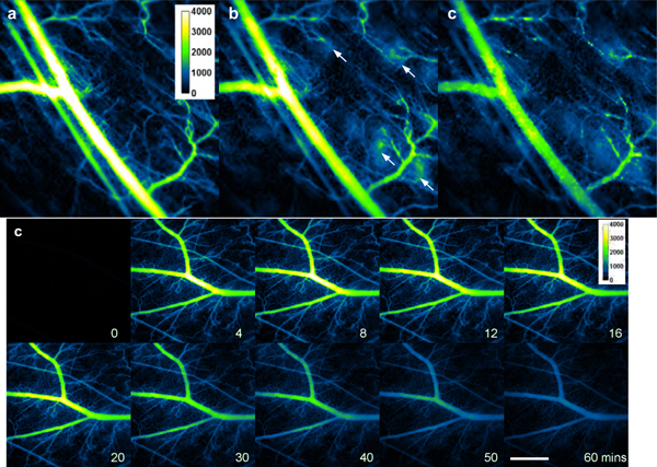
Intravital NIR fluorescent images of mouse skin microvasculature in the window chamber in response to second injection of μNETs (7 days after first injection), (a-d) Fluorescent images acquired at 3, 13, 30, and 60 minutes for one mouse. (e-f) Fluorescent images at 12 and 10 minutes post-injection, respectively, for a second and third mouse. In Panels (b), (e) and (f), bright spots are visible in the regions around the tips of the capillaries (white arrows). In Panels (c) and (d), there are large and bright particles on the vessel wall. White bar represents 1 mm and applies to all panels. NIR images are falsely-colored. NETs were fabricated from bovine erythrocytes.