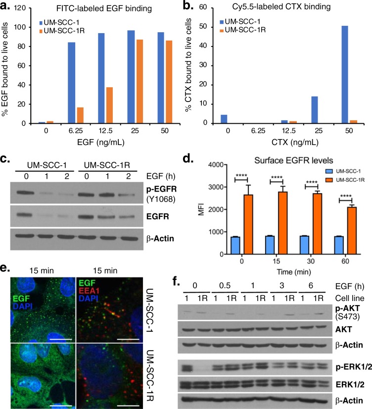
Fig 2. Effect of G33S and N56K mutants on EGF or CTX binding and EGFR activation and degradation.
(a-b) FITC-labeled EGF and Cy5.5-labeled CTX in UM-SCC-1 vs.–SCC-1R (G33K-N56 mut) assessed by flow cytometry after 30 min of incubation with various concentrations of EGF or CTX. (c) Phospho- and total EGFR levels at indicated times of incubation with saturating EGF (60 ng/mL). (d) Surface levels of EGFR in cells stimulated with 60 ng/mL EGF in unpermeabilized/unfixed cells, by flow cytometry using a secondary goat anti-rabbit Alexa-Fluor 488 antibody. (e) Mapping of Alexa-Fluor 488-EGF conjugate shows internalization (green dotted lipid rafts) and co-localization with early endosome in UM-SCC-1 but not UM-SCC-1R. Scale bar represents 10 μM. (f) Consistently increased phospho-AKT but overall comparable (p)ERK1/2 levels in the mutant (1R = UM-SCC-1R) vs. parental (1 = UM-SCC-1) cells. Blots for (p)ERK1/2 were generated on a separate gel with its own β-actin loading control.

