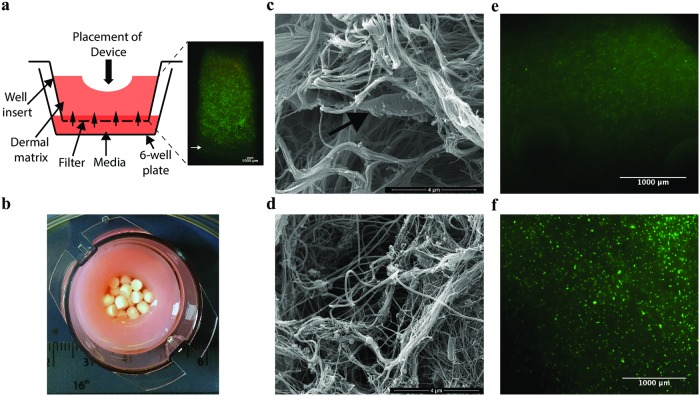
Fig 1. The IVM model system is such that actives can be added to the surface of the model or the media and the matrix is comparable to that of soft tissue.
(a) Schematic of the IVM model system and live dead staining of the fibroblasts in cross-section through the model after two weeks maintenance in tissue culture. Green spots indicate live cells, red indicates dead cells, as cells are viable in this image there are very few red spots. (b) Image of the IVM model from above showing the central void packed with CSB with measurements for scale. (c) SEM image of the IVM model with a fibroblast indicated in the collagen matrix by a white arrow (d) compared to DFAN, a sample of debrided tissue. Micrographs of live dead stained sections of representative control models which were not inoculated with bacteria and to which (e) unloaded CSB or (f) gentamicin loaded CSB were added in parallel to inoculated models. There was no evidence of toxicity of the gentamicin or calcium sulfate.

