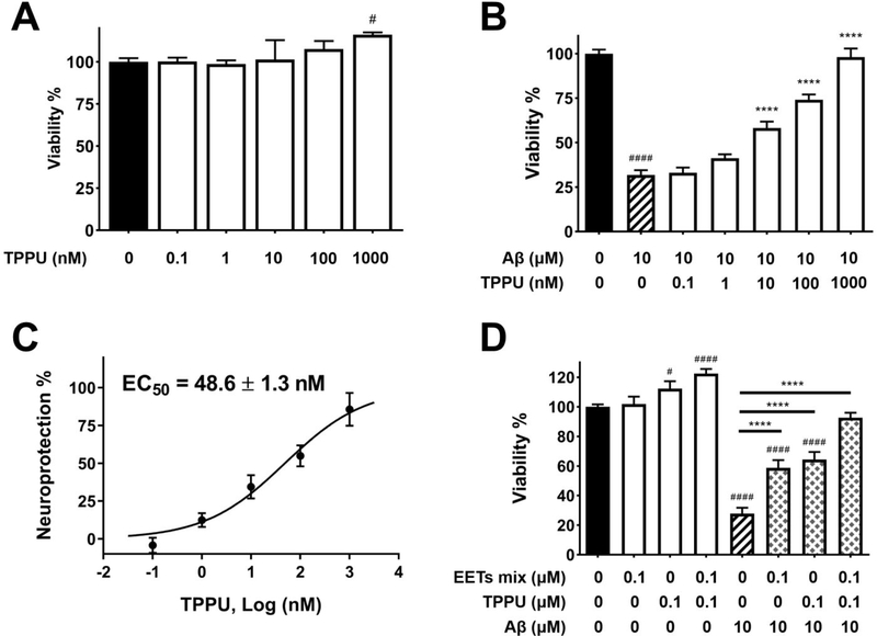
Figure 5.
TPPU inhibited Aβ42 neurotoxicity in SH-SY5Y cells. Cell viability was determined with the MTS assay. Data were the mean of duplicate of six independent experiments with ± SEM (n = 6). Data were analyzed by one-way ANOVA with Tukey’s multiple comparison test. #p < 0.05, ####p < 0.0001 relative to the vehicle control; ****p < 0.0001 relative to the 10 μM Aβ42 treatment. (A) Cytotoxicity assessment of TPPU in SH-SY5Y cells. Cells were treated with varying concentrations of TPPU or 0.2% PEG 400 vehicle and incubated for 72 h. (B) Cells were pretreated with varying concentrations of TPPU or 0.2% PEG 400 vehicle for 1 h followed by 10 μM Aβ42 treatment for 72 h. (C) TPPU inhibited neurotoxicity induced by 10 μM Aβ42 with an EC50 value of 48.6 nM. The results were normalized as the percentage of the neuroprotective activity relative to the control (100%) and the 10 μM Aβ42 treatment (0%). Neuroprotection curve was analyzed by four-parameter regression. (D) Cotreatment of EETs with TPPU enhanced neuroprotection.

