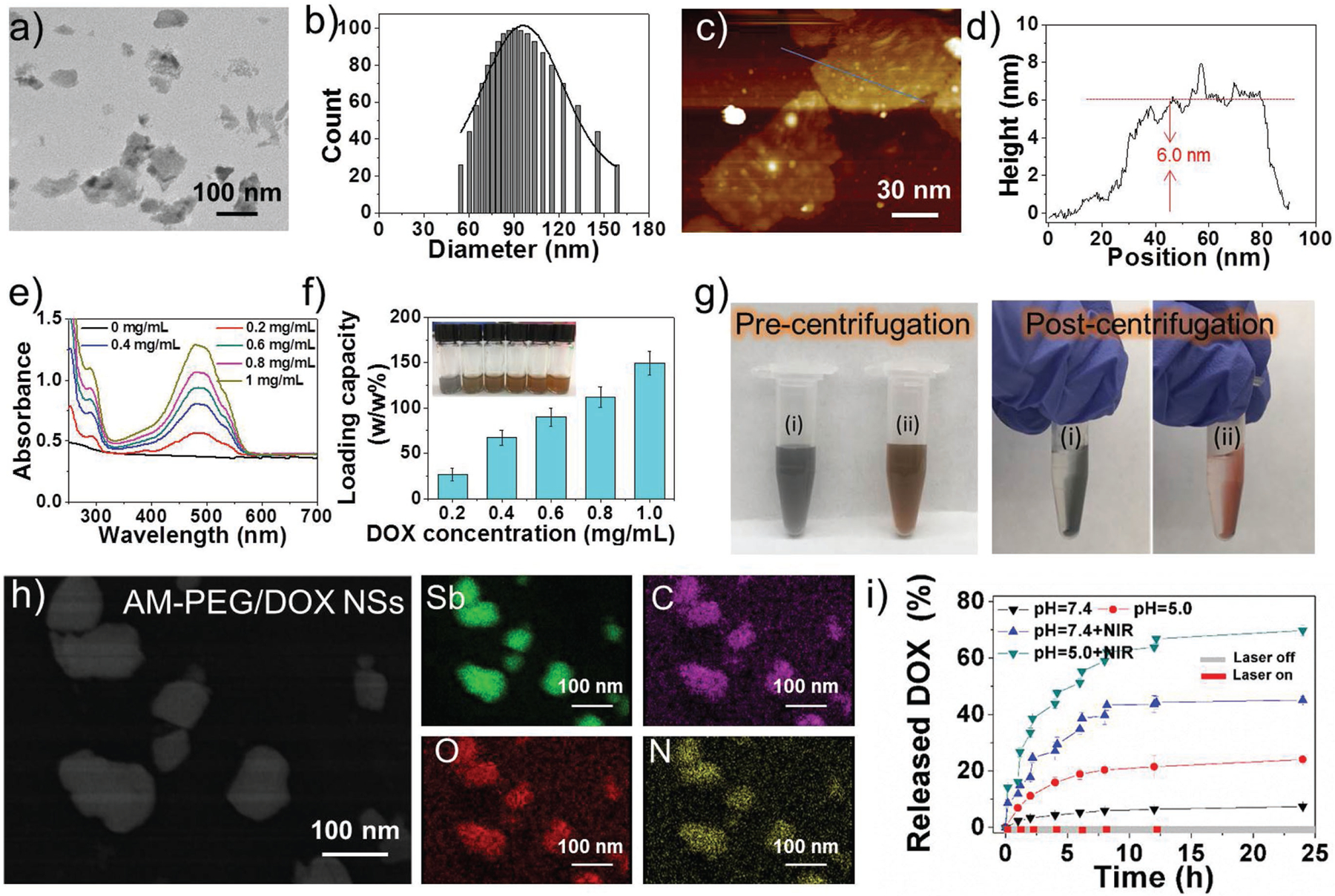
Figure 1.

Characterization of the AM-based NSs. a) TEM image, b) size distribution, c) AFM image, and d) thickness measured from (c) of the 2D AM-PEG NSs. e) UV–vis–NIR absorbance of AM-PEG/DOX NSs at different DOX feeding concentrations after the removal of excess-free DOX. f) DOX loading capacities on AM-PEG NSs (w/w%) with increasing DOX feeding concentrations. g) Photographs of (i) AM-PEG NSs and (ii) AM-PEG/DOX NSs solution before and after centrifugation. h) STEM-EDC mapping images of AM-PEG/DOX NSs. i) Release profiles of DOX at different pHs with or without 808-nm NIR laser (0.8 W cm−2).
