Figure.
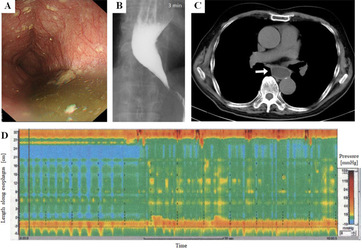
Images of the diagnostic examinations in a patient with achalasia. (A) Upper gastrointestinal endoscopy showing intra-esophageal retention of food debris within the extended esophagus. (B) Barium swallow test showing a dilated esophagus with a “bird’s beak” appearance. (C) Chest CT showing a dilated esophagus with the retained liquid inside (white arrow). (D) High-resolution manometry showing aperistalsis and absent esophageal pressurization, which is compatible with the criteria of Type I achalasia in the Chicago classification.

