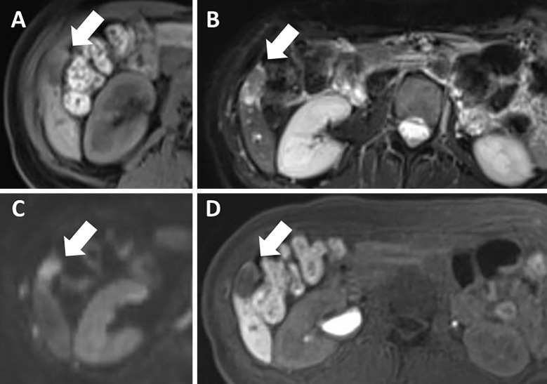
Figure 2.
On ethoxybenzyl-magnetic resonance imaging performed two months before the patient’s admission, the tumor (arrows) had not changed in size and was isointense compared with the normal liver on T1-weighted imaging (T1WI) (A). It was also highly heterogeneous on T2-weighted imaging (T2WI) (B) and diffusion-weighted imaging (DWI) (C); there was no uptake in the hepatobiliary phase (D).

