FIGURE 2.
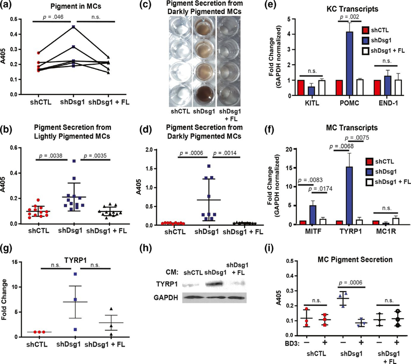
Exposure to conditioned media from Dsg1-deficient KCs increases MC pigmentation and pigment secretion. (a) Seven days of exposure to conditioned media from Dsg1-deficient (shDsg1) KCs increased MC pigment content (read as absorbance at 405 nm following lysis with NaOH) compared with conditioned media from control (CTL) KCs or media from KCs infected with shDsg1 plus silencing-resistant Dsg1-Flag (shDsg1 + FL) (N = 5 MC:KC-conditioned media pairs, plotted as spaghetti plots to represent each individual experiment). (b) Quantification of pigment secretion into the media from MCs from lightly pigmented donors (Caucasian, Asian, Hispanic, unknown) treated for 7 days with conditioned media from KCs infected with shCTL, shDsg1, shDsg1 + FL constructs (N = 13 MC:KC-conditioned media pairs). (c) Example of visual appearance of pigment secretion into the media (concentrated by centrifugation) from MCs from 4 individual darkly pigmented donors (African American) treated for 7 days with the indicated conditioned media. (d) Quantification of pigment secretion into the media from MCs from darkly pigmented donors (African American) treated for 7 days with the indicated conditioned media (N = 9 MC:KC-conditioned media pairs). Treatment with conditioned media from KCs depleted of Dsg1 significantly increased pigment secretion into the media compared with conditioned media from shCTL or shDsg1 + FL-infected KCs. For panels A, B, and D, data were analyzed using one-way ANOVA followed by Tukey’s post hoc analysis. (e) Pro-opiomelanocortin (POMC) mRNA was increased in shDsg1-infected KCs compared with shCTL and shDsg1 + FL-infected KCs, while KITL and END-1 mRNA levels were not significantly changed between groups (N = 3 independent experiments, KCs were harvested on day 3 after being induced to differentiate in high calcium-containing medium). (f) Transcripts involved in pigment production in MCs (microphthalmia-associated transcription factor [MITF] and tyrosinase-related protein 1 [TYRP1] were upregulated while melanocortin 1 receptor [MC1R]) were not significantly changed in MCs treated with conditioned media from shDsg1-infected KCs compared to MCs treated with conditioned media from shCTL or shDsg1 + FL-infected KCs (N = 3 independent experiments). For panels E and F, gene expression was normalized to GAPDH across samples and then reported as fold change compared with shCTL. Reported p values are results of one-way ANOVA of the fold change data followed by Tukey’s post hoc analysis. (g) Quantification of immunoblots performed after 3 MC isolates were treated with conditioned media from shCTL, shDsg1, or shDsg1 + FL-infected KCs. TYRP1 protein increased in all 3 MC isolates treated with conditioned media from shDsg1-infected KCs (did not reach the level of significance). (h) An example TYRP1 immunoblot is shown. GAPDH serves as a protein loading control. (i) Recombinant human beta-defensin 3 (BD3, 100 nM) in conditioned media (7 days) from KCs depleted of Dsg1 inhibits MC pigment secretion (N = 3, data were analyzed using one-way ANOVA followed by Holm-Sidak’s post hoc analysis for comparison between drug-treated and drug-untreated samples for each condition). Graphical data in B, D, G, and I are represented as mean and standard deviation since each individual data point represents a single experimental replicate. qPCR data in E and F are represented as mean and SEM

