Fig 4.

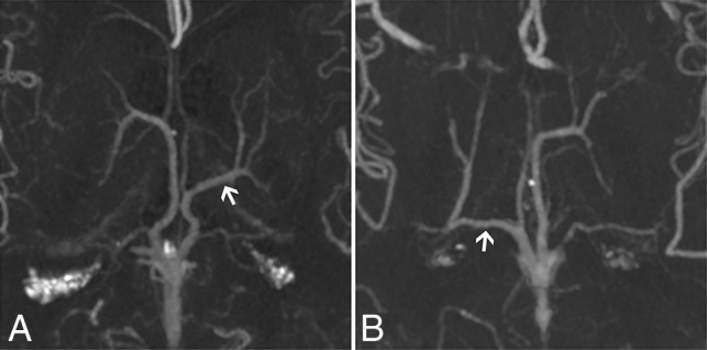
CTA MIP axial cross-sections of the ICV branching patterns with an LDV and an absent thalamostriate vein. Arrows indicate LDVs. A, The suprathalamic LDV (type 2; left hemisphere). B, The retrothalamic LDV (type 3; right hemisphere).

CTA MIP axial cross-sections of the ICV branching patterns with an LDV and an absent thalamostriate vein. Arrows indicate LDVs. A, The suprathalamic LDV (type 2; left hemisphere). B, The retrothalamic LDV (type 3; right hemisphere).