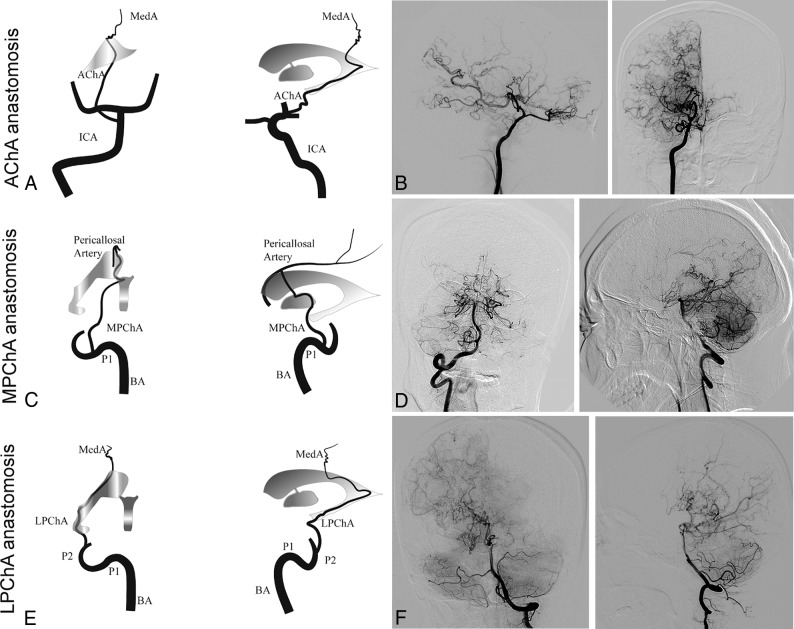
Fig 1.
Schematic illustrations and angiographic findings from representative cases of each subtype of choroidal collateral anastomosis. A and B, Anterior-posterior and lateral right carotid artery angiograms show a dilated anterior choroidal artery extending beyond the lateral ventricle to the cortex (arrows). C and D, Anterior-posterior and lateral right vertebral artery angiograms show a medial posterior choroidal artery extending beyond the level of the pericallosal artery (arrows) to the corpus callosum. E and F, Anterior-posterior and lateral left vertebral artery angiograms show a lateral posterior choroidal artery extending beyond the body of the lateral ventricle to the cortex (arrows). MedA indicates the medullary artery; P1 and P2, the proximal portion of posterior cerebral artery; BA, the basilar artery.

