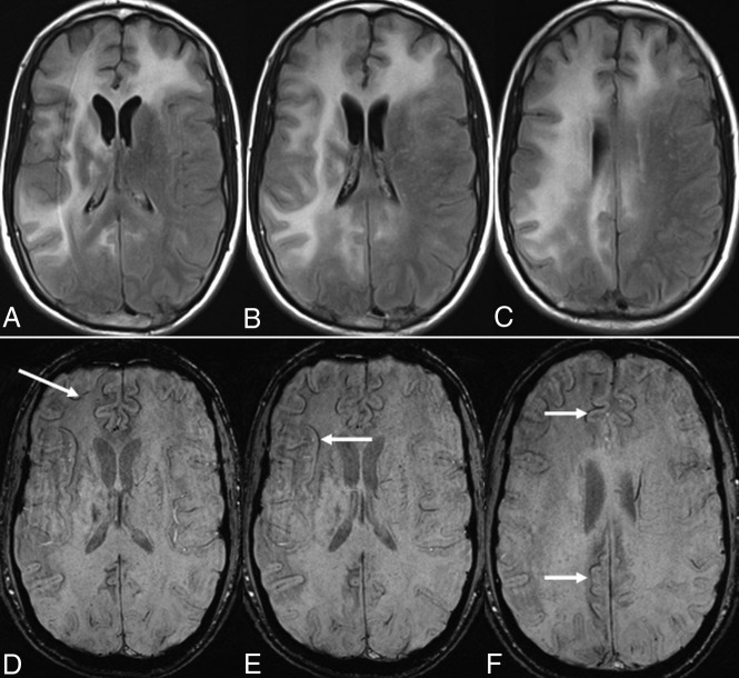
Fig 2.
On axial FLAIR images (A–C), multifocal PML lesions are observed in both frontal lobes, the capsula interna and externa, and the right parietal lobe. A linear, relatively thin hypointense rim is observed on the cortical side of the lesions on axial SWI (D–F) in a long-term survivor (9 years, still alive).

