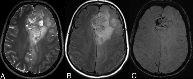
Fig 2.
A 54-year-old woman with a left frontal lobe oligodendroglioma, IDH-mutant and 1p/19q-codeleted, showing characteristic imaging features. A and B, T2WI and FLAIR demonstrate a heterogeneous and poorly marginated mass with significant cortical infiltration and no T2-FLAIR mismatch sign. C, T2*WI shows regions of striking susceptibility blooming.

