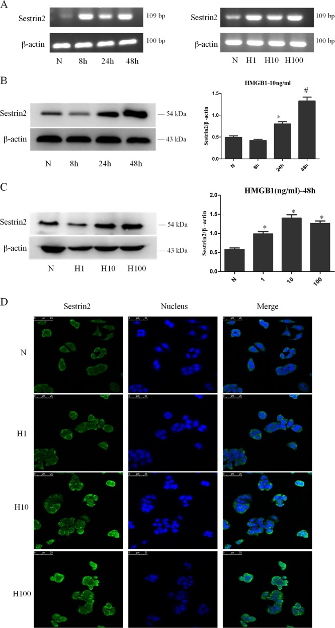
Fig. 2. HMGB1 upregulated the expression of SESN2 in DC2.4 cells.
a SESN2 mRNA expression were analyzed by PCR after DC2.4 cells stimulated with HMGB1 (10 ng/ml) for different intervals (0, 8, 24, and 48 h), or with various dosages of HMGB1 (1, 10, and 100 ng/ml) for 48 h. β-actin served as the internal standard. b, c Protein levels of SESN2 were analyzed by Western blotting after DC2.4 cells stimulated with 10 ng/ml HMGB1 for different time points or at various concentrations for 48 h. β-actin served as the internal standard, n = 3 per group. d Laser scanning confocal microscopy was employed to observe the expression of SESN2 protein in DC2.4 cells after HMGB1 treatment at various concentrations for 48 h, with FITC labeled SESN2 protein (green) and DAPI stained nucleus (blue) (×600), scale bar = 25 μm, n = 4 per group. Data were presented as mean ± SD of at least three independent experiments. Statistical significance: *P < 0.05, #P < 0.01 versus the control group.

