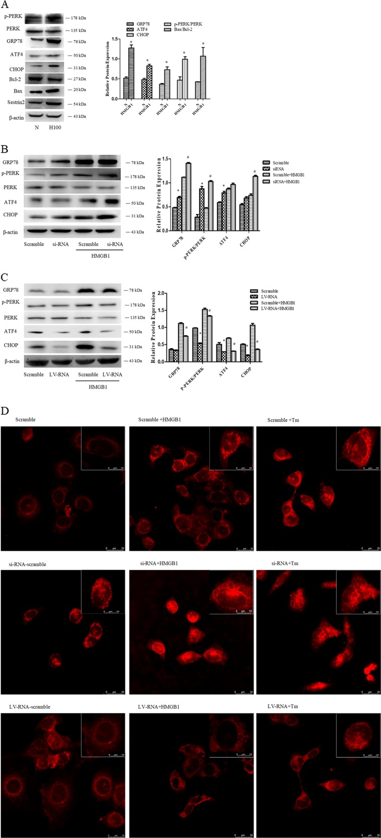
Fig. 6. SESN2 protected DC2.4 cells against excessive ERS response induced by HMGB1.

a Expressions of GRP78, p-PERK, PERK, ATF4, CHOP, Bcl-2, and Bax were determined by Western blotting after treatment with 100 ng/ml HMGB1 for 48 h. b, c Lentiviral vectors (SESN2 knockdown and SESN2 overexpression) were transduced DC2.4 cells, and scramble cells were transduced with blank vectors. After treated with 100 ng/ml HMGB1 for 48 h, expressions of GRP78, p-PERK, PERK, ATF4, and CHOP were determined by Western blot analysis. d The morphologic alteration of ER in each cell was evaluated by immunofluorescence staining and the extent of the ER morphologic change was compared with the scramble controls (×600, ×1200), scale bar = 25 μm, scale bar = 10 μm, n = 4 per group. ER was detected with ER-tracker red. β-actin served as the internal standard. Data were presented as the mean ± SD, n = 3 per group. Statistical significance: *P < 0.05 versus the control group.
