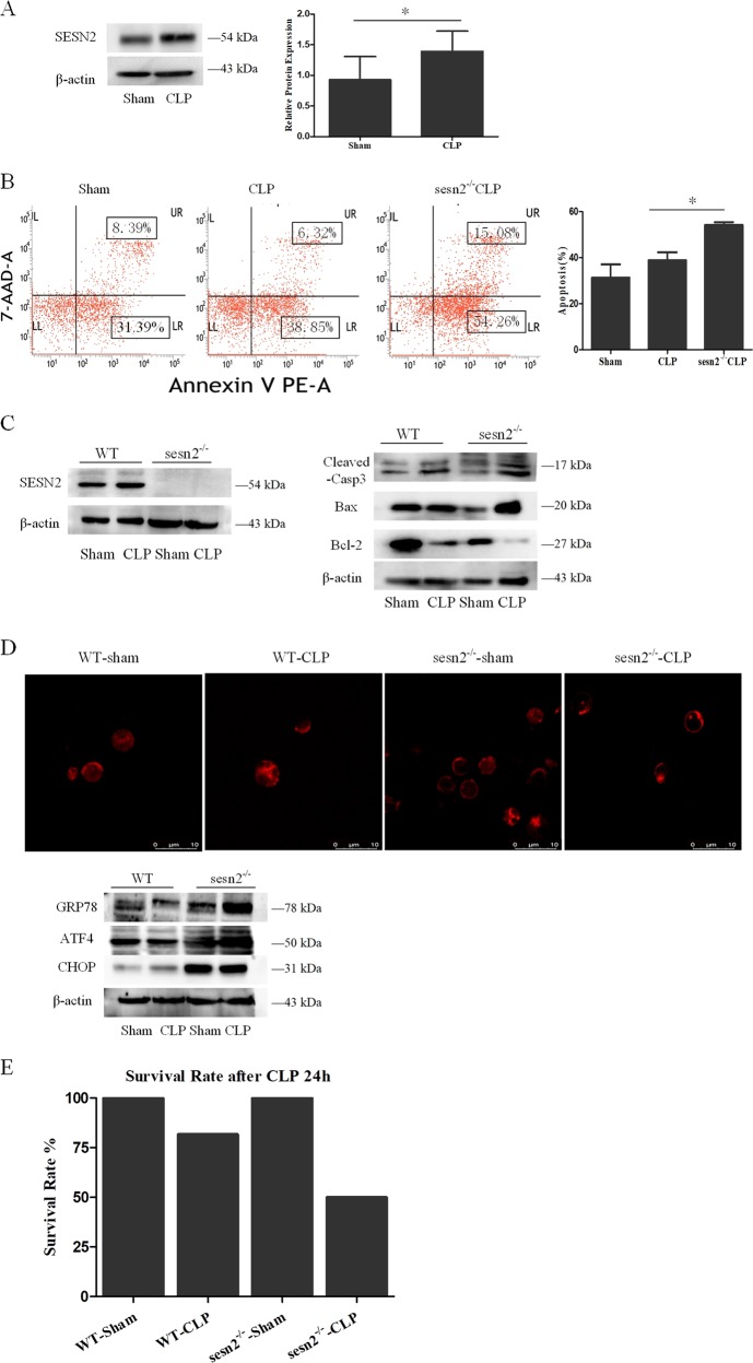
Fig. 8. The protective effect of SESN2 on DCs in CLP-induced septic mice.
Mice underwent a sham procedure or CLP. a SESN2 expression was determined by Western blotting at 24 h after after CLP. b PE-Annexin-V and 7-AAD were used to stain DCs and subjected to flow cytometry to assess cell apoptosis at 24 h after CLP in vivo. c Expressions of cleaved-caspase-3, Bcl-2, and Bax were measured as described in the section of methods after CLP procedure. d In the sesn2−/− CLP group, the morphologic alteration of ER in each cell was evaluated by immunofluorescence staining and the extent of the ER morphologic change was compared with the WT CLP group (×1200). ER was detected with ER-tracker red. Levels of GRP78, ATF4, and CHOP were assessed by Western blotting. β-actin served as the internal standard. e The 24-h survival rate of mice in the sesn2−/− CLP group was markedly lower than that in the WT CLP group. Data of three independent experiments were presented as the mean ± SD, n = 6 per sham group, n = 11–13 per CLP group. Statistical significance: *P < 0.05 for in vivo comparison of the sesn2−/− CLP group versus the WT CLP group.

