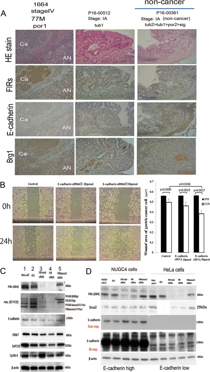
Fig. 6. E-cadherin siRNA promotedg astric cancer cells migration in the wound-healing assay.
a E-cadherin expression decreased, whereas FIR family and BRG1 expressions increased in cancer tissues (Ca) relative to those in adjacent normal tissues revealed by immunohistochemical staining. Case numbers 2832, 1621, and 1664 are listed in Suppl. Table S4. In the non-invasive early stage (IA) of differentiated cancers, the expressions of E-cadherin and FBW7 were decreased. Case number P16-00361 are listed in Suppl. Table S8. b Migration of E-cadherin siRNAs transfected into NUGC4 cells and corresponding control cells was measured by wound-healing assay. P < 0.01 was obtained by Student’s t-test. c FIR family and related protein expressions were examined aftertreatment of HeLa cells withthe FIRsiRNAs. GL2 siRNA was transfected as the negativecontrol. After 48 h of transfection, whole-cell extracts were analyzed by western blotting. Three types of FIR siRNAs were transfected into HeLa cells. Lane 2 is GL2 siRNA control transfection, lane 3 is 20 pmol of total FIR siRNA transfection, lane 4 is 20 pmol of FIRsiRNA transfection, and lane 5 is 20 pmolof FIRΔexon2 siRNA transfection. d E-cadherin expression in HeLa cells. The level of E-cadherin expression was much higher in NUGC4 than that of HeLa cells. Three types of FIR siRNAs were transfected into NUGC4 and HeLa cells. GL2 siRNA is internal control. 20 pmol of total FIR siRNA, 20 pmol of FIR siRNA, and 20 pmol of FIRΔexon2 siRNA transfection were performed. After 48 h of transfection, whole-cell extracts were analyzed by western blotting.

