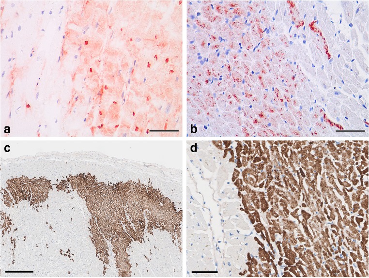
Fig. 6.
Immunostaining of early myocardial infarction. Positive staining for fibronectin (a) and C5b-9 (b) in irreversibly injured cardiomyocytes. Scale bars = 50 μm. Courtesy from Aljakna et al., Int J Legal Med, 2018; acute myocardial infarction in papillary muscle immunostained with C4d antibody (brown). Low power view, bar = 0.25 mm, highlights exact delineation of necrotic areas (geographic zones, and multifocal cells) (c); Higher magnification, bar = 50 μm, shows abrupt border between vital tissue and necrotic area (d)

