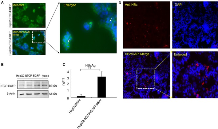
Figure 2.

NTCP‐EGFP mediates infection by HBV in HepG2 cells. (A) HepG2‐NTCP‐EGFP cell lines were observed under the fluorescence microscope. The white arrow indicates the EGFP signal on the membrane. (B) Total protein was isolated from HepG2‐NTCP‐EGFP cells, and the expression of NTCP was identified by Western blot (C) HepG2 cells or HepG2‐NTCP‐EGFP cells were infected with HBV (150 Geq/cell) for 24 h. Cells were fixed and supernatant was collected at day 12 d.p.i. Secreted HBsAg was detected by TRIFA. (D) HBcAg expression was determined by indirect immunofluorescence. (Results are the means ± SD of three repeats, with differences assessed using Student's t test. **P < .01. Scale bar = 100 μm)
